8 Free Heart Anatomy transparent PNG images
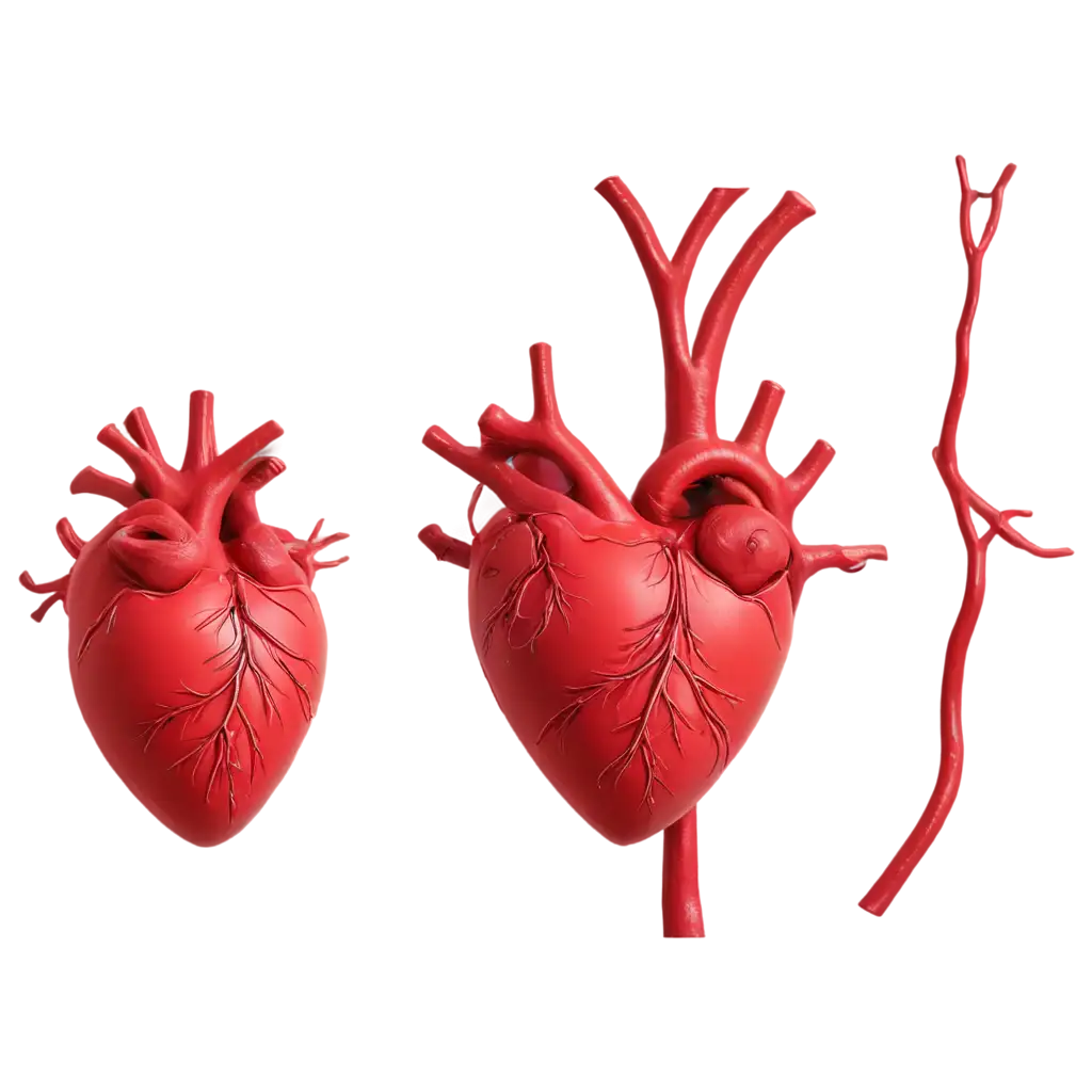
Explore our comprehensive Heart Anatomy image collection, featuring 8 free AI-generated images that bring medical visualization to life. From detailed anatomical stock photos to dynamic 3D renderings, vectors, and professional medical illustrations, our diverse collection serves both educational and professional needs. Each high-resolution image is available for immediate download, and with our innovative 'open in editor' feature, you can fine-tune the prompts to generate variations that perfectly match your requirements.



Related Tags
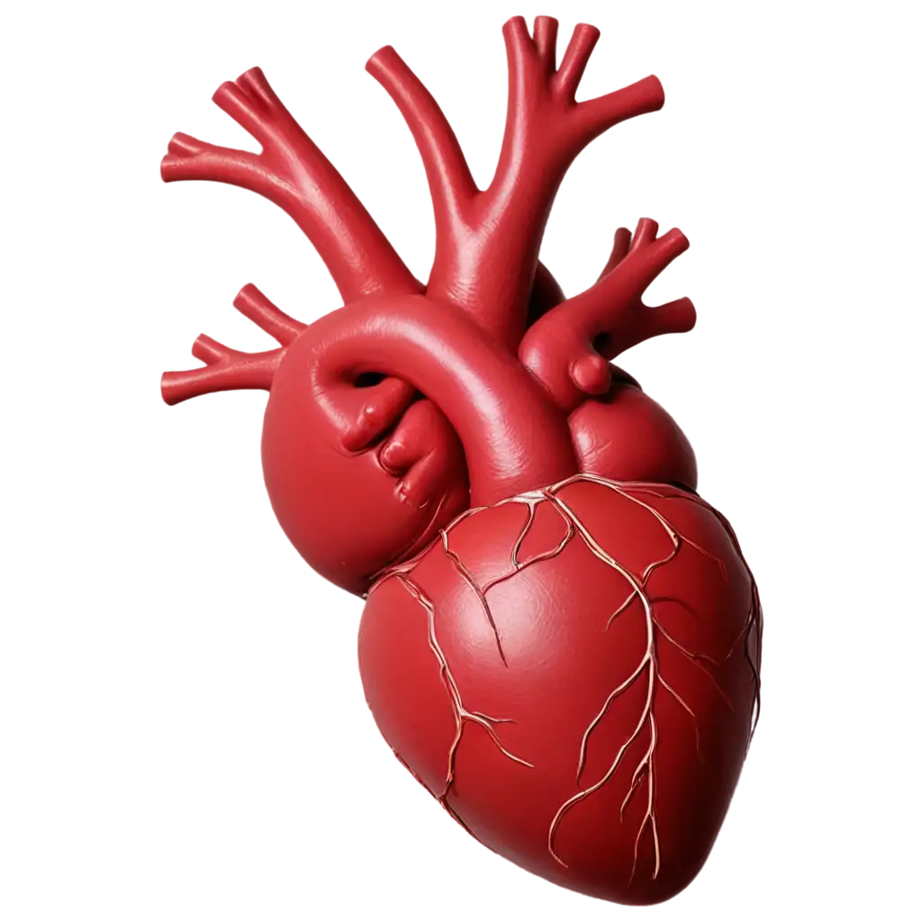
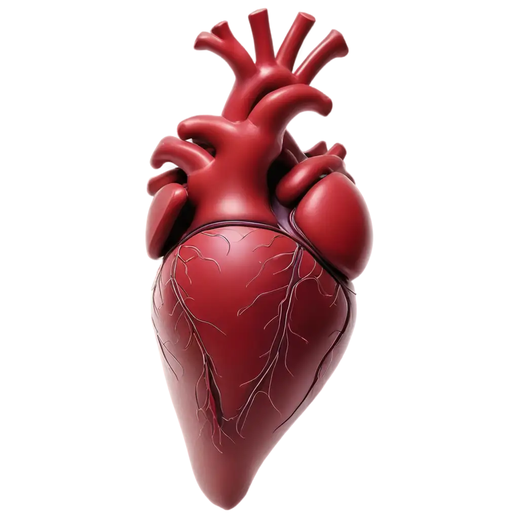
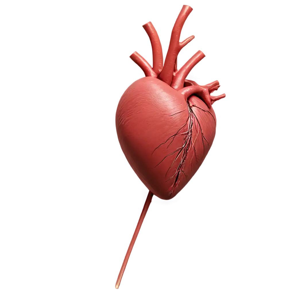
Heart anatomy visualization has evolved significantly with the advent of AI technology. Our collection showcases intricate details of cardiac structures, from the four chambers (atria and ventricles) to the complex network of blood vessels, valves, and surrounding tissues. These AI-generated images provide unprecedented clarity in depicting both external and internal cardiac anatomy, making them invaluable for medical education, professional presentations, and patient education. The anatomical accuracy is maintained through careful prompt engineering and validation against medical references, ensuring that each image serves as a reliable visual resource.
Understanding Heart Anatomy Through AI-Generated Imagery
AI-generated heart anatomy images serve multiple purposes across the medical field. In educational settings, these visualizations help students grasp complex anatomical concepts through clear, three-dimensional representations. Healthcare professionals use them for patient communication, making it easier to explain conditions and procedures. The images are particularly valuable in medical presentations, publications, and educational materials, where their high resolution and customizable nature allow for precise illustration of specific cardiac features. The variety of styles available - from photorealistic to schematic representations - enables users to choose the most appropriate visualization for their specific needs.
Applications of Heart Anatomy Images in Medical Education and Communication
The process of generating accurate heart anatomy images involves sophisticated AI algorithms trained on extensive medical imaging datasets. Key considerations include proper anatomical proportions, accurate coloration of different tissues, and precise representation of cardiac structures. Users can achieve optimal results by incorporating specific medical terminology in their prompts, such as 'anterior interventricular sulcus' or 'coronary sinus.' The 'open in editor' feature allows for real-time adjustments to details like viewpoint, cross-sectional planes, and tissue transparency, enabling the creation of customized anatomical visualizations that meet specific educational or professional requirements.
Creating Accurate Heart Anatomy Visualizations with AI
The field of AI-generated medical imagery is rapidly evolving, with emerging trends pointing toward even more sophisticated visualization capabilities. Future developments include integration with augmented reality for interactive 3D viewing, real-time pathology visualization, and dynamic representation of cardiac function. Machine learning algorithms are becoming increasingly adept at generating highly detailed anatomical structures, promising even more accurate and versatile heart anatomy visualizations. These advancements will further enhance the utility of AI-generated images in medical education, clinical communication, and research applications.
Future Trends in AI-Generated Medical Anatomy Visualization